
Anatomy by Planes
Complex Joints Motions
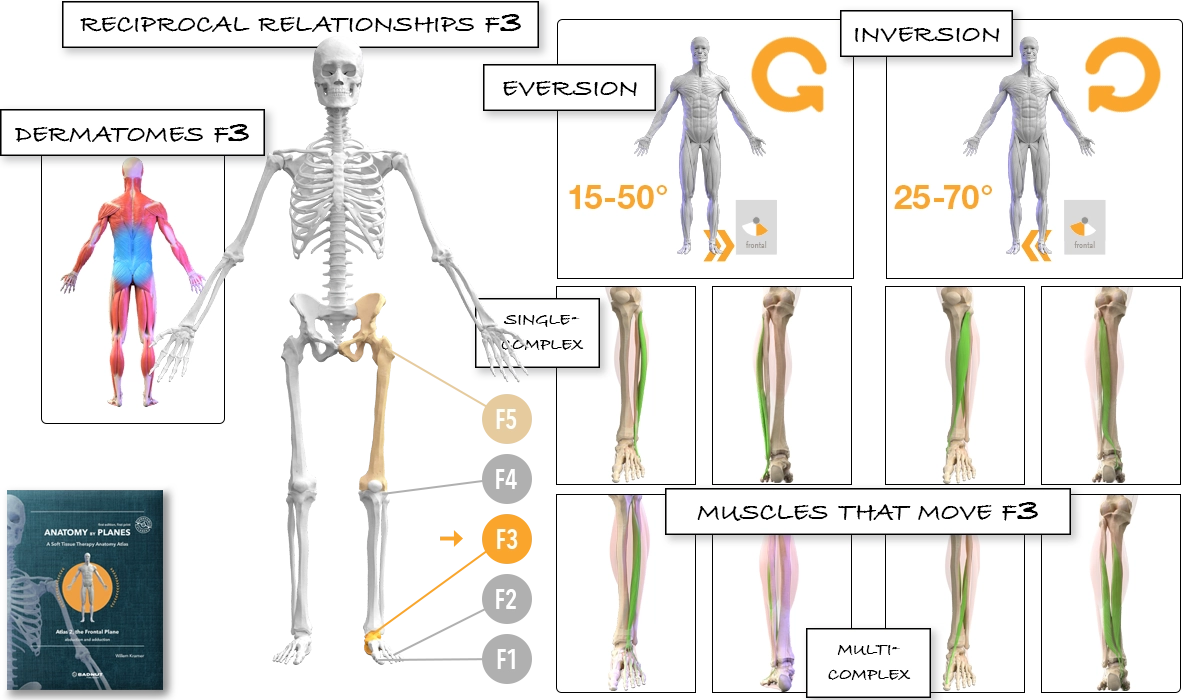
Frontal plane.
Frontal ankle, F3.
Talocalcenal joint.
Transverse tarsal joints.
15 - 50 degrees eversion.
25 - 70 degrees inversion.
Indirect Motion Reciprocal Relationships # passive complex
T11 - S2.
None.
Frontal hip, F5.
Frontal knee, F4. #
Frontal ankle, F3. ←
Frontal lesser toes, F2. #
Frontal big toe, F1. #
Muscles Single-Complex Depth of Muscle
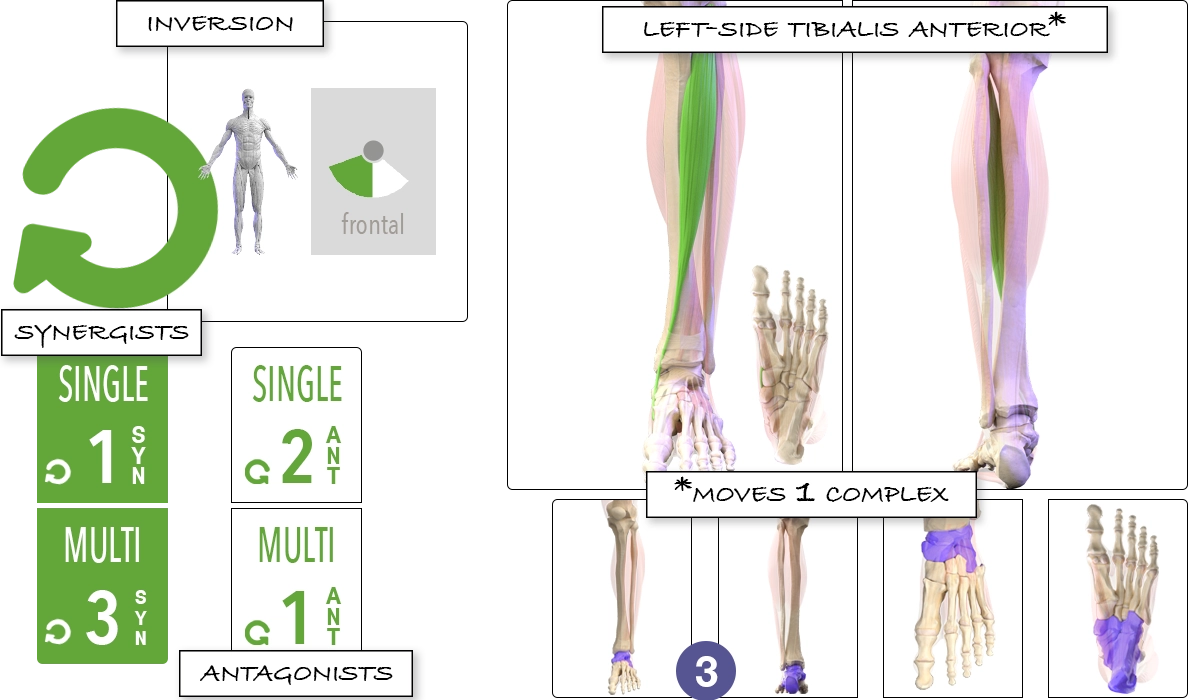
Inversion (and adduction).
Tibialis anterior.
Ankle. ←
Level 1, just under the skin.
Single-Complex Antagonists (2) Multi-Complex Synergists (3) Multi-Complex Antagonists (1)
Tibialis posterior.
Peroneus longus & brevis.
Peroneus tertius.
Extensor hallucis longus.
Flexor digitorum longus.
Flexor hallucis longus.
Extensor digitorum longus.
Traditional Anatomy
Proximal attachments
The tibialis anterior connects with the upper half to two-thirds of the lateral tibia, from its lateral condyle on down, the interosseous membrane, the fascia cruris, and the intermuscular septum between it and the extensor digitorum longus.
Distal attachment
The tibialis anterior connects with the medial and plantar surfaces of the medial cuneiform and the medial surface of the base of the first metatarsal bone.
Location
Located just lateral to the anterior border of the tibia, directly under the skin. It has a long tendon that starts around the lower third of the tibia. Passing underneath the superior and inferior extensor retinaculae, the tendon crosses the distal tibia, moving from the lateral to the medial side of the leg.
Innervation
Deep peroneal nerve.
Vascular Supply
Medial and anterior branches of the anterior tibial artery and the anterior tibial recurrent artery.
The tendon is supplied by the anterior medial malleolar, the dorsalis pedis artery, the medial tarsal arteries, and the medial malleolar and calcaneal branches of the posterior tibial artery.
Motion
Dorsiflexion.
Inversion.