

In this series, I share one complex per post. Starting with the sagittal big toe, working my way up to the sagittal thumb, before doing the same with the complexes of the frontal (aka, coronal) and transverse (aka, horizontal) planes, respectively. The first post (read HERE) in the series explains single- and multi-complex muscles, introduces complexes as I use them in Anatomy by Planes, and explains why complexes make it easier and more effective to resolve pain complaints through treatment and exercise. Enjoy!
The Sagittal Big Toe
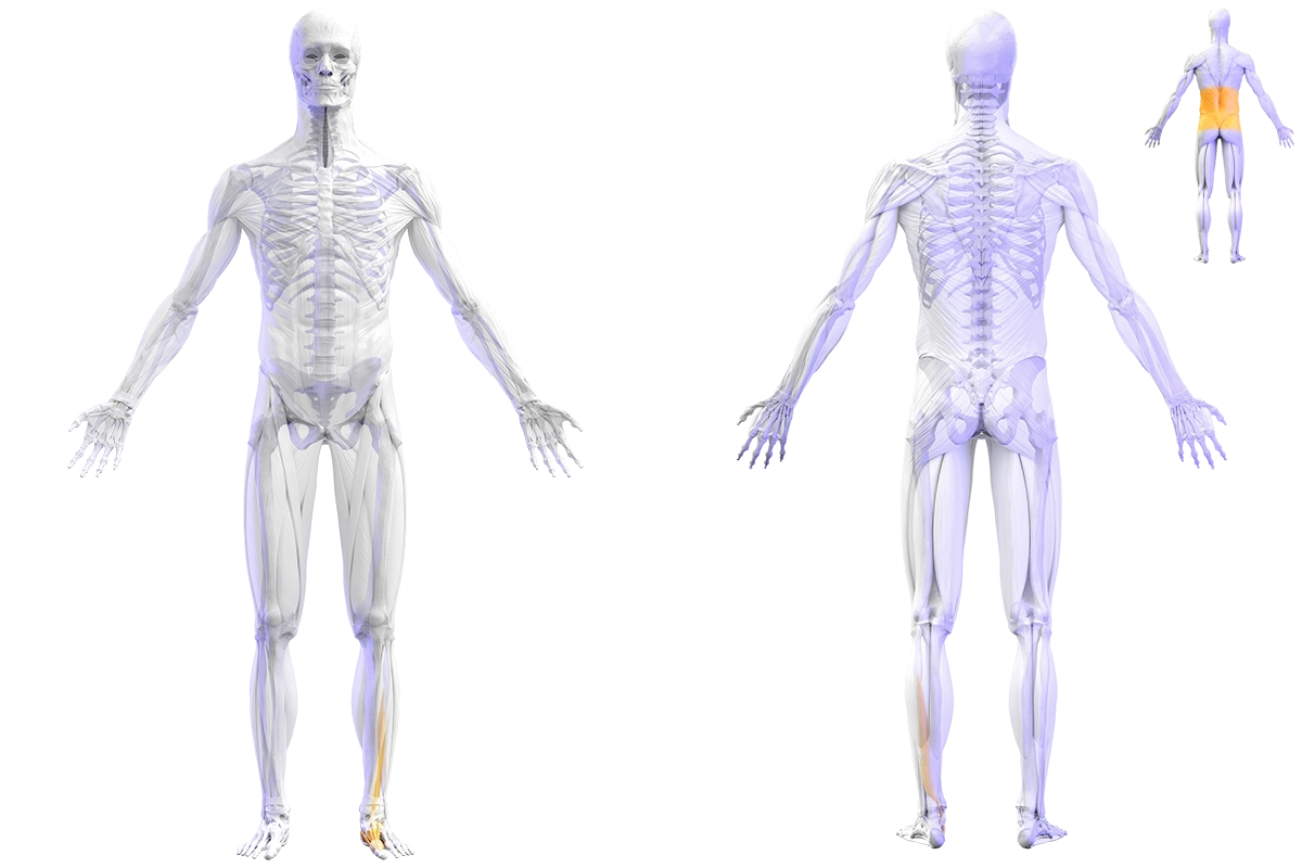
The sagittal big toe is the first complex of the sagittal plane. It serves as a key anchor and pivot point during functional movement. The orange colored parts of the integumentary, skeletal, and muscular systems (see below) belong to it and are involved in its flexion and extension motions.
The skin, joints, and muscles of the sagittal big toe are reciprocally related to each other (intra-complex). The complex as a whole is reciprocally related to its neighboring complexes (inter-complex). Through these relationships, the sagittal big toe can contribute to and help resolve a wide range of pain conditions, injuries, and dysfunction.

Associated Pain, Injuries, and Dysfunction
- Hallux limitus [1, 2]
- Hallux rigidus [1, 2]
- Turf toe [3, 4, 5]
- Sesamoid pathology [3, 4]
- Transfer metatarsalgia [6, 7, 8]
- Plantar plate tears (2nd MTP) [9, 10]
- Hammertoe and crossover toe [10, 11]
- Morton's neuroma [12]
- Plantar fasciitis / plantar fasciopathy [13, 14]
- Lateral ankle sprain [15, 16]
- Chronic ankle instability [15, 16]
- Limited ankle dorsiflexion (functional or structural) [17, 18]
- Posterior ankle impingement syndrome [19, 20]
- Shin splints (medial tibial stress syndrome) [21, 22]
- Flexor hallucis longus tendinopathy [20]
- Achilles tendinopathy [23]
- Runner's knee (patellofemoral pain syndrome) [24, 25, 26]
- Jumper's knee (patellar tendinopathy) [27, 28, 29, 30]
- Hip flexor strain [31, 32]
- Lower back pain [31, 32]
Treating and Exercising Complex by Complex
For each of these conditions, the sagittal big toe is either the primary site of pathology or a contributing factor through the reciprocal relationships above. If a client visits you with one of these problems, treating and exercising the skin, joints, and muscles of the sagittal big toe can help them feel better; sometimes a lot, sometimes a little.
Overview Sagittal Big Toe Complex (flexion-extension)
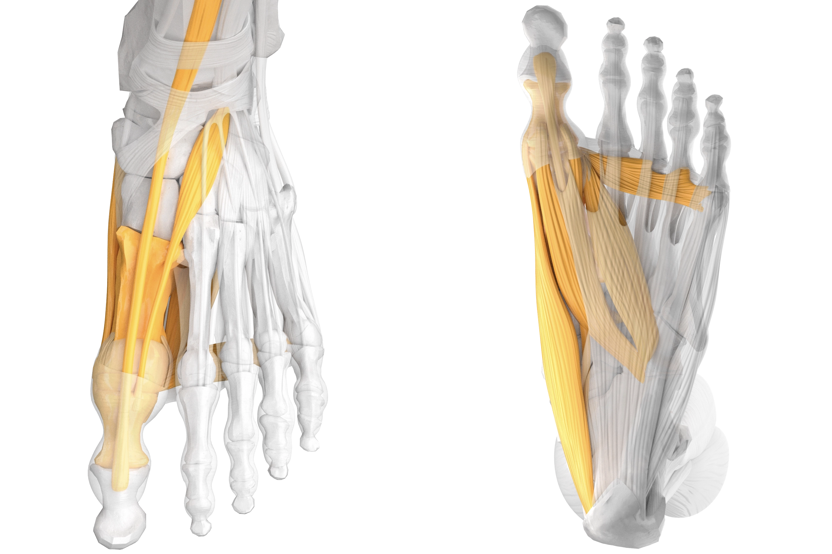
Skin, Joints, and Muscles of the Sagittal Big Toe
ROM (range of motion): 15-40 degrees flexion and 55-95 degrees extension.
Skin: over the single-complex muscles and dermatomes T11-S2 (paraspinal posterior torso). The range includes both somatic and ANS source segments — the two are integrated at the segmental level (see van Cranenburgh, Segmentale verschijnselen en van Zutphen, Nederlands leerboek der fysische therapie in engine zin).
Joints: first metatarsophalangeal joint (aka first MTP). The first MTP joint consists of the first metatarsal, proximal phalanx, and the joint capsules, ligaments, and plantar plate complex that hold the bones together. Additionally, some literature considers the medial and lateral sesamoids (embedded in the distal tendons of the flexor hallucis brevis) part of the MTP joint, despite a distinct anatomical and biomechanical separation.
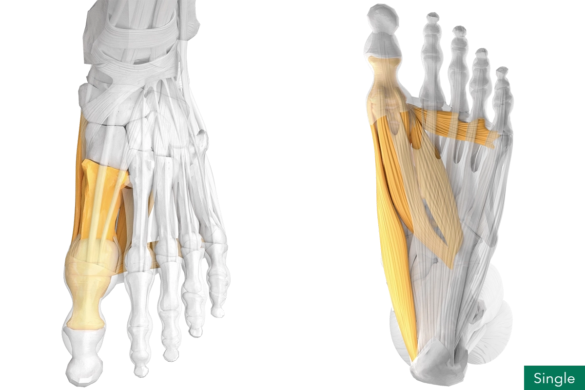
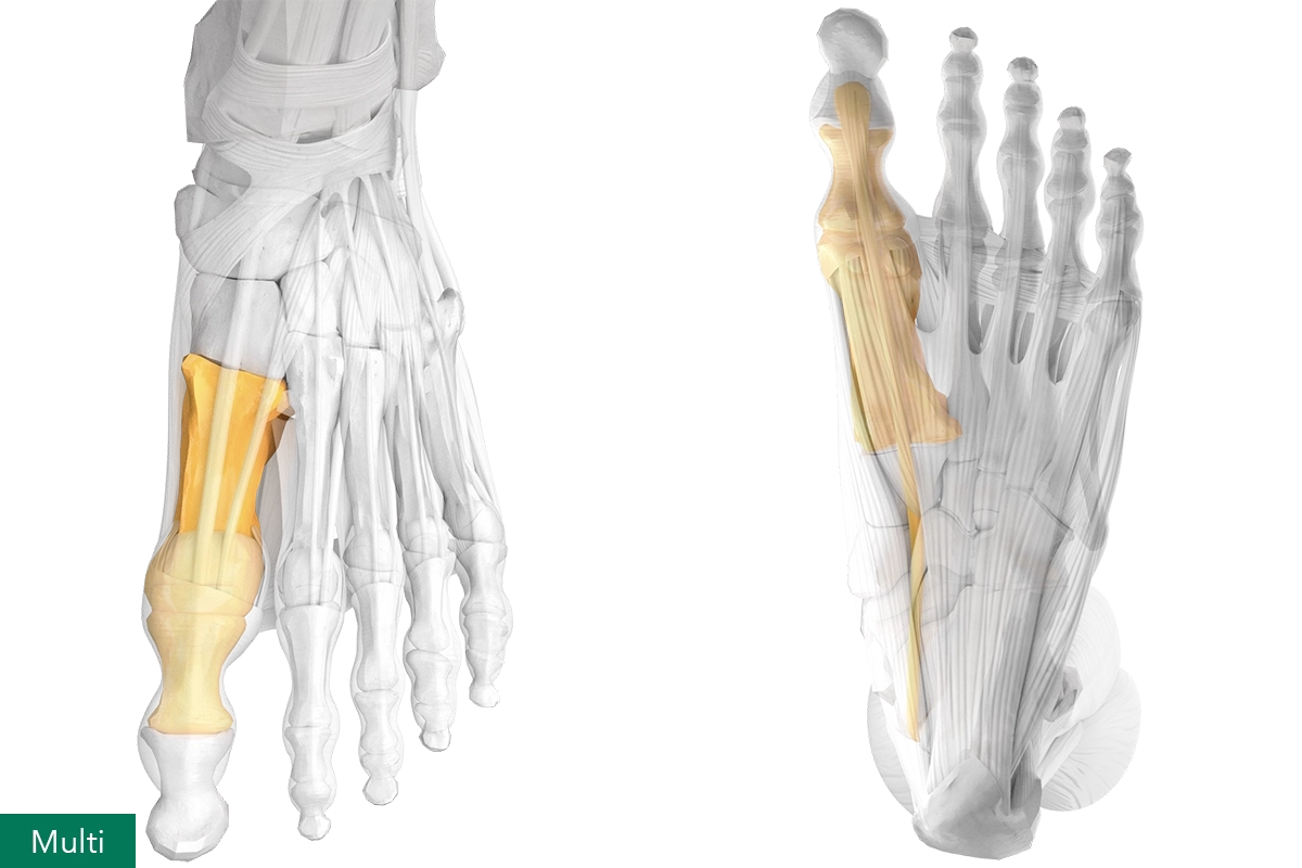
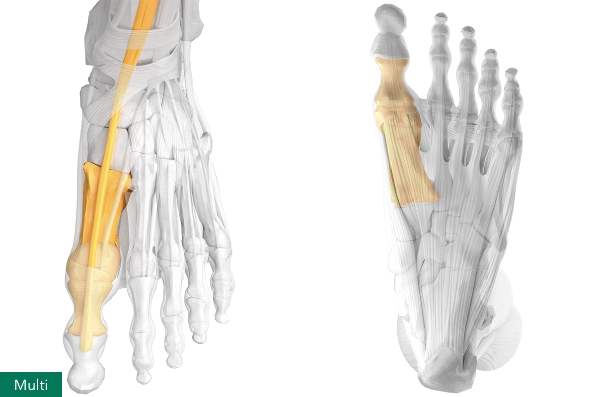
Muscles: abductor hallucis, adductor hallucis (both heads), extensor hallucis brevis, extensor hallucis longus, flexor hallucis brevis, flexor hallucis longus.
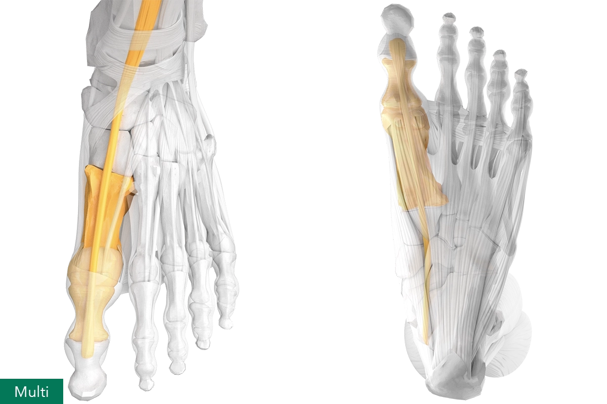
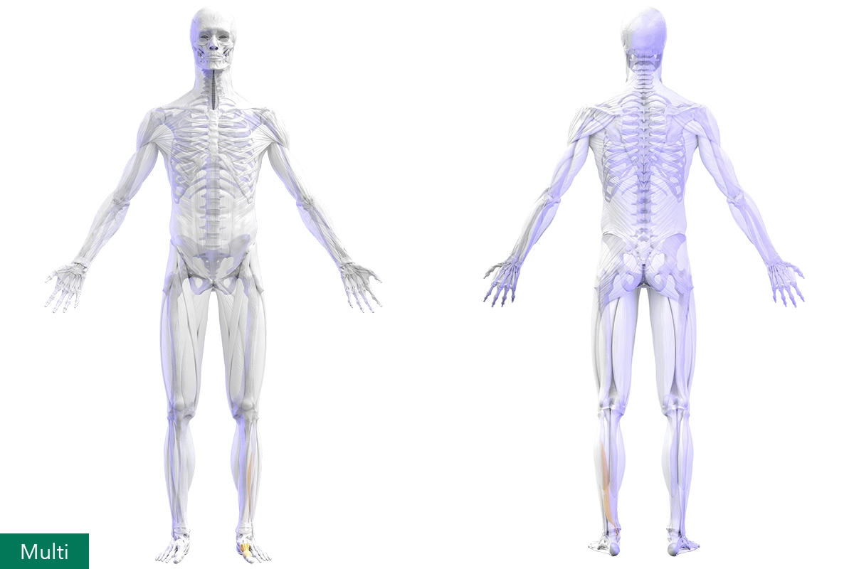
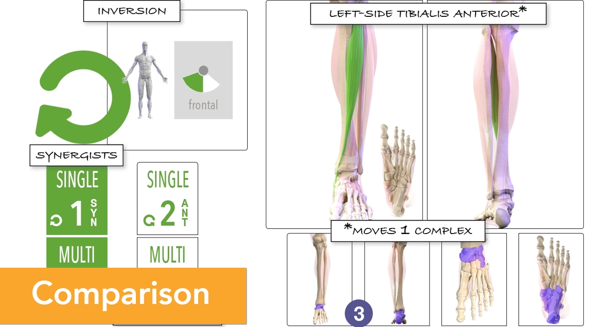
Single-Complex Muscles vs Multi-Complex Muscles


Single-Complex Muscles: abductor hallucis, adductor hallucis (both heads), extensor hallucis brevis, flexor hallucis brevis.
Multi-Complex Muscles: extensor hallucis longus, flexor hallucis longus.
Single-Complex Flexor Muscles vs Multi-Complex Flexor Muscles

Single-Complex Flexors: abductor hallucis, adductor hallucis (both heads), flexor hallucis brevis.
Multi-Complex Flexors: flexor hallucis longus.
Single-Complex Extensor Muscles vs Multi-Complex Extensor Muscles


Single-Complex Extensors: extensor hallucis brevis.
Multi-Complex Extensors: extensor hallucis longus.
References Associated Pain, Injuries, and Dysfunction of the Sagittal Big Toe
[1] Patel J, Swords M. Hallux Rigidus. In: StatPearls [Internet]. Treasure
Island (FL): StatPearls Publishing; updated November 22, 2023. [PMID: 32310479]
[2] Tovaruela-Carrión N, Becerro-de-Bengoa-Vallejo R, Losa-Iglesias ME,
López-López D, Gómez-Salgado J, Bayod-López J. Exploring the Association of
Hallux Limitus with Baropodometric Gait Pattern Changes. Bioengineering.
2025;12(3):316. [doi: 10.3390/bioengineering12030316]
[3] Mason LW, Molloy AP. Turf Toe and Disorders of the Sesamoid Complex. Clin
Sports Med. 2015;34(4):725–739. [doi: 10.1016/j.csm.2015.06.008]
[4] Frimenko RE, Lievers WB, Coughlin MJ, Anderson RB, Crandall JR, Kent RW.
Etiology and biomechanics of first metatarsophalangeal joint sprains (turf
toe) in athletes. Crit Rev Biomed Eng. 2012;40(1):43–61. [doi: 10.1615/CritRevBiomedEng.v40.i1.30]
[5] Gupta A, Singh PK, Xu AL, Bronheim RS, McDaniel CM, Aiyer AA. Turf Toe
Injuries in the Athlete: an Updated Review of Treatment Options,
Rehabilitation Protocols, and Return-to-Play Outcomes. Curr Rev Musculoskelet
Med. 2023;16(11):563–574. [doi: 10.1007/s12178-023-09870-y]
[6] Chong XL, Drittenbass L, Dubois-Ferriere V, Assal M. Iatrogenic transfer
metatarsalgia after hallux valgus surgery: a comprehensive treatment
algorithm. EFORT Open Rev. 2022;7(9):618–627. [doi: 10.1530/EOR-22-0043]
[7] Slullitel G, López V, Calvi JP, Seletti M, Bartolucci C, Pinton G. Effect
of First Ray Insufficiency and Metatarsal Index on Metatarsalgia in Hallux
Valgus. Foot Ankle Int. 2016;37(3):300–306. [doi: 10.1177/1071100715615323]
[8] Walker AK, Heinen RM. The Role of First Ray Insufficiency in the
Development of Metatarsalgia. Foot Ankle Clin. 2019;24(4):641–648. [doi: 10.1016/j.fcl.2019.07.001]
[9] Doty JF, Coughlin MJ. Metatarsophalangeal joint instability of the lesser
toes and plantar plate deficiency. J Am Acad Orthop Surg. 2014;22(4):235–245. [doi: 10.5435/JAAOS-22-04-235]
[10] Akoh CC, Phisitkul P. Plantar Plate Injury and Angular Toe Deformity.
Foot Ankle Clin. 2018;23(4):703–713. [doi: 10.1016/j.fcl.2018.07.010]
[11] Bouché RT, Heit EJ. Combined plantar plate and hammertoe repair with
flexor digitorum longus tendon transfer for chronic, severe sagittal plane
instability of the lesser metatarsophalangeal joints. J Foot Ankle Surg.
2008;47(2):125–137. [doi: 10.1053/j.jfas.2007.12.008]
[12] Gougoulias N, Lampridis V, Sakellariou A. Morton's interdigital neuroma:
instructional review. EFORT Open Rev. 2019;4(1):14–24. [doi: 10.1302/2058-5241.4.180025]
[13] Bolgla LA, Malone TR. Plantar Fasciitis and the Windlass Mechanism: A
Biomechanical Link to Clinical Practice. J Athl Train. 2004;39(1):77–82. [PMID: 15085218]
[14] Moisan G, Chicoine D, McBride S, Isabelle PL, Gómez-Carrión Á, Farahpour
N. First metatarsophalangeal joint dorsiflexion resistance in individuals
with plantar fasciopathy. Foot (Edinb). 2024;60:101140. [PMID: 39142073]
[15] Fraser JJ, Koldenhoven RM, Jaffri AH, Park JS, Saliba SF, Hart JM, Hertel
J. Foot impairments contribute to functional limitation in individuals with
ankle sprain and chronic ankle instability. Knee Surg Sports Traumatol
Arthrosc. 2020;28(5):1600–1610. [doi: 10.1007/s00167-018-5028-x]
[16] Alshahrani MS, Reddy RS, Alshahrani A, Gautam AP, Alsubaie SF. Exploring
the interplay between ankle muscle strength, postural control, and pain
intensity in chronic ankle instability: A comprehensive analysis. Heliyon.
2024;10(5):e27374. [doi: 10.1016/j.heliyon.2024.e27374]
[17] Martinez-Rico M, Gijon-Nogueron G, Ortega-Avila AB, Roche-Seruendo LE,
Climent-Pedrosa A, Sanchis-Sales E, Deschamps K. Effect of Custom-Made Foot
Orthotics on Multi-Segment Foot Kinematics and Kinetics in Individuals with
Structural Hallux Limitus. Sensors (Basel). 2024;24(19):6430. [doi: 10.3390/s24196430]
[18] Tovaruela-Carrión N, Becerro-de-Bengoa-Vallejo R, Losa-Iglesias ME,
López-López D, Gómez-Salgado J, Bayod-López J. Hallux Limitus: Exploring the
Variability in Lower Limb Symmetry and Its Connection to Gait Parameters — A
Case–Control Study. Bioengineering. 2025;12(3):298. [doi: 10.3390/bioengineering12030298]
[19] Maquirriain J. Posterior ankle impingement syndrome. J Am Acad Orthop
Surg. 2005;13(6):365–371. [doi: 10.5435/00124635-200510000-00001]
[20] Hamilton WG, Geppert MJ, Thompson FM. Pain in the posterior aspect of
the ankle in dancers. Differential diagnosis and operative treatment. J Bone
Joint Surg Am. 1996;78(10):1491–1500. [doi: 10.2106/00004623-199610000-00006]
[21] Reshef N, Guelich DR. Medial tibial stress syndrome. Clin Sports Med.
2012;31(2):273–290. [doi: 10.1016/j.csm.2011.09.008]
[22] Saad MA, Jamal JM, Aldhafiri AT, Alkandari SA. Medial Tibial Stress
Syndrome: A Scoping Review of Epidemiology, Biomechanics, and Risk Factors.
Cureus. 2025;17(3):e81463. [doi: 10.7759/cureus.81463]
[23] Wirth SH, Andronic O, Aregger F, Jungwirth-Weinberger A, Jentzsch T,
Hecker A. Flexor hallucis longus hypertrophy secondary to Achilles tendon
tendinopathy: an MRI-based case–control study. Eur J Orthop Surg Traumatol.
2021;31(7):1387–1393. [doi: 10.1007/s00590-021-02891-8]
[24] Davis IS, Powers CM. Patellofemoral Pain Syndrome: Proximal, Distal, and
Local Factors — An International Research Retreat. J Orthop Sports Phys Ther.
2010;40(3):A1–A48. [doi: 10.2519/jospt.2010.0302]
[25] Barton CJ, Levinger P, Webster KE, Menz HB. Kinematics associated with
foot pronation in individuals with patellofemoral pain syndrome: a
case-control study. J Foot Ankle Res. 2011;4(Suppl 1):O4. [doi: 10.1186/1757-1146-4-S1-O4]
[26] Kaya D, Atay OA, Callaghan MJ, Cil A, Caglar O, Citaker S, Yuksel I,
Doral MN. Hallux valgus in patients with patellofemoral pain syndrome. Knee
Surg Sports Traumatol Arthrosc. 2009;17(11):1364–1367. [doi: 10.1007/s00167-009-0775-3]
[27] Backman LJ, Danielson P. Low range of ankle dorsiflexion predisposes for
patellar tendinopathy in junior elite basketball players: a 1-year
prospective study. Am J Sports Med. 2011;39(12):2626–2633. [doi: 10.1177/0363546511420552]
[28] Tayfur A, Haque A, Salles JI, Malliaras P, Screen H, Morrissey D. Are
Landing Patterns in Jumping Athletes Associated with Patellar Tendinopathy?
A Systematic Review with Evidence Gap Map and Meta-analysis. Sports Med.
2022;52(1):123–137. [doi: 10.1007/s40279-021-01550-6]
[29] Martinez AF, Scattone Silva R, Paschoal BLF, Souza LLA, Serrão FV.
Association of Ankle Dorsiflexion and Landing Forces in Jumping Athletes.
Sports Health. 2022;14(6):932–937. [doi: 10.1177/19417381211063456]
[30] Mendonça LD, Ocarino JM, Bittencourt NFN, Macedo LG, Fonseca ST.
Association of Hip and Foot Factors With Patellar Tendinopathy (Jumper's
Knee) in Athletes. J Orthop Sports Phys Ther. 2018;48(9):676–684. [doi: 10.2519/jospt.2018.7426]
[31] Dananberg HJ. Gait style as an etiology to chronic postural pain. Part
I. Functional hallux limitus. J Am Podiatr Med Assoc. 1993;83(8):433–441. [PMID: 8409128]
[32] Dananberg HJ. Gait style as an etiology to chronic postural pain. Part
II. Postural compensatory process. J Am Podiatr Med Assoc.
1993;83(11):615–624. [PMID: 8295677]
References General Anatomy
[33] Kelikian AS, Sarrafian SK. Sarrafian's Anatomy of the Foot and Ankle:
Descriptive, Topographic, Functional. 3rd ed. Philadelphia, PA: Lippincott
Williams & Wilkins; 2011. [WorldCat: ISBN 9780781797504]
[34] Standring S, ed. Gray's Anatomy: The Anatomical Basis of Clinical
Practice. 42nd ed. Amsterdam: Elsevier; 2021. [WorldCat: ISBN 9780702077050]
[35] Kapandji AI. The Physiology of the Joints. Volume Two, Lower Limbs. 5th
ed. New York: Churchill Livingstone; 1987. [WorldCat: ISBN 9780443036187]
[36] Kendall FP, McCreary EK, Provance PG, Rodgers MM, Romani WA. Muscles:
Testing and Function with Posture and Pain. 5th ed. Baltimore, MD: Lippincott
Williams & Wilkins; 2005. [WorldCat: ISBN 9780781747806]
[37] Draves DJ. Anatomy of the Lower Extremity. Baltimore, MD: Williams &
Wilkins; 1986. [WorldCat: ISBN 9780683026511]
[38] Travell JG, Simons DG. Myofascial Pain and Dysfunction: The Trigger
Point Manual. Volume 2: The Lower Extremities. Baltimore, MD: Williams &
Wilkins; 1992. [WorldCat: ISBN 9780683083675]
References Anatomy of the Sagittal Big Toe
[39] Glasoe WM, Yack HJ, Saltzman CL. Anatomy and biomechanics of the first
ray. Phys Ther. 1999;79(9):854–859. [doi: 10.1093/ptj/79.9.854]
[40] Davies MB, Dalal S. Gross anatomy of the interphalangeal joint of the
great toe: implications for excision of plantar capsular accessory ossicles.
Clin Anat. 2005;18(4):239–244. [doi: 10.1002/ca.20069]
[41] Hicks JH. The mechanics of the foot. II. The plantar aponeurosis and
the arch. J Anat. 1954;88(1):25–30. [PMID: 13129168]
[42] Brenner E. Insertion of the abductor hallucis muscle in feet with and
without hallux valgus. Anat Rec. 1999;254(3):429–434. [doi: 10.1002/(SICI)1097-0185(19990301)254:3<429::AID-AR14>3.0.CO;2-5]
[43] Akita K, Niiro N, Murakami G, Sato T. First dorsal interosseous muscle
of the foot and its innervation. Clin Anat. 1999;12(1):12–15. [doi: 10.1002/(SICI)1098-2353(1999)12:1<12::AID-CA2>3.0.CO;2-K]
[44] Akita K, Sakamoto H, Sato T. Lateromedial and dorsoplantar borders
among supplying areas of the nerves innervating the intrinsic muscles of the
foot. Anat Rec. 1999;255(4):465–470. [doi: 10.1002/(SICI)1097-0185(19990801)255:4<465::AID-AR12>3.0.CO;2-Z]
[45] Kalin PJ, Hirsch BE. The origins and function of the interosseous
muscles of the foot. J Anat. 1987;152:83–91. [PMID: 3654378]
References Skin, Dermatomes, and Segmental SNS-ANS Anatomy
[46] Lee MW, McPhee RW, Stringer MD. An evidence-based approach to human
dermatomes. Clin Anat. 2008;21(5):363–373. [doi: 10.1002/ca.20636]
[47] Bernards ATM. Fysiologie en pathofysiologie van nocisensoriek. In: van
Zutphen HCF, van Sambeek HWR, Oostendorp RAB, eds. Nederlands leerboek der
fysische therapie in engere zin, Deel I. 4th ed. Utrecht: Wetenschappelijke
uitgeverij Bunge; 1991. [WorldCat: title search]
[48] van Cranenburgh B. Segmentale verschijnselen: een bijdrage aan
diagnostiek en therapie. 2nd ed. Houten: Bohn Stafleu van Loghum; 2000. [WorldCat: ISBN 9789031343188]


